Renal Ultrasound: Complete Guide to Prep, Procedure & Results
Ever wondered what’s going on inside your kidneys? A renal ultrasound is a simple, painless way to get a peek. Think of it like a super-powered camera that uses sound waves to create detailed pictures of your kidneys. This guide will explain everything about this amazing imaging technique, from what to expect before, during, and after the scan, to understanding what the results mean. We’ll cover the basics in plain English, so you can feel comfortable and informed every step of the way, helping you understand your kidney health better.
Understanding Renal Ultrasound: A Detailed Guide
Let’s explore renal ultrasounds – a non-invasive method doctors use to examine your kidneys. It’s a straightforward procedure that can reveal valuable information about your kidney health. Consider it a high-tech sonar for your kidneys, providing detailed images to aid doctors in assessing their size, shape, and condition.
What is a Renal Ultrasound Used For?
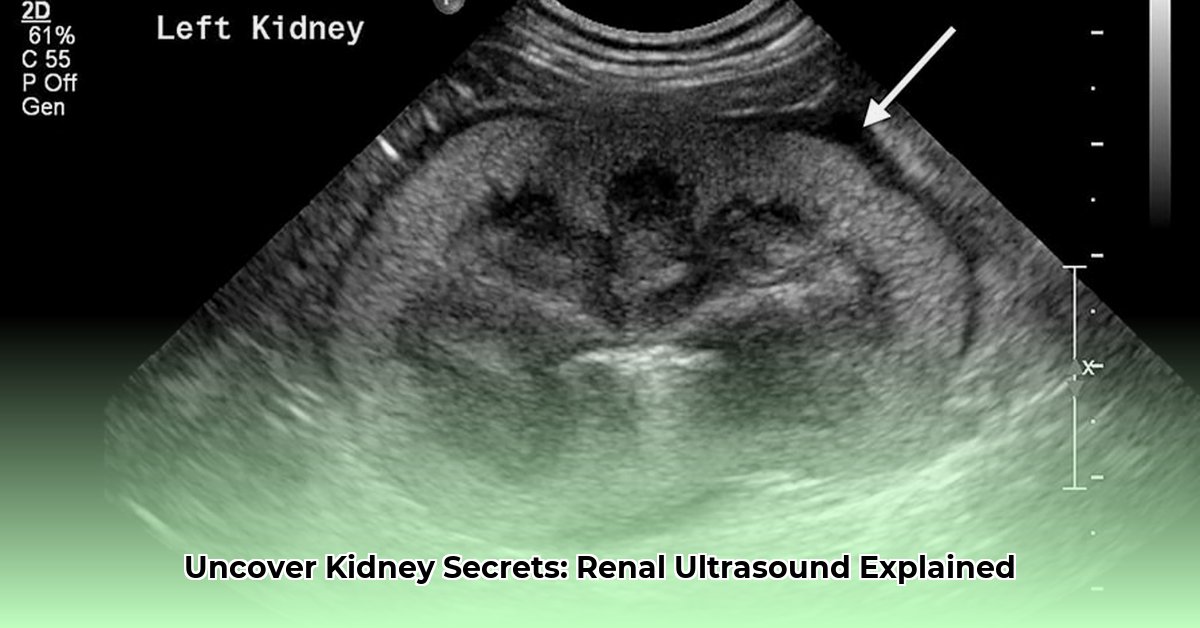
A renal ultrasound, also known as a kidney ultrasound, uses sound waves to create images of your kidneys. Unlike X-rays, it doesn’t use radiation, making it a safe imaging choice. The ultrasound helps healthcare providers visualize the size, shape, and location of your kidneys. It can also detect abnormalities like cysts, stones, and blockages, aiding in the diagnosis of various kidney conditions. A complete ultrasound often includes images of the bladder and surrounding structures.
How Does a Renal Ultrasound Work?
Imagine sound waves, like tiny echoes, bouncing off your kidneys. A special device called a transducer emits these inaudible sound waves. These waves travel through your body and reflect back from your kidneys and surrounding tissues. The transducer captures these reflected waves and converts them into a real-time image displayed on a monitor. This image allows the doctor to assess the size, shape, and structure of your kidneys, as well as identify any potential abnormalities.
How is a Renal Ultrasound Performed?
Understanding the procedure beforehand can help ease any anxiety. Here’s a step-by-step guide to what you can expect during a renal ultrasound:
Step 1: Preparation
In most cases, you can eat and drink normally before a renal ultrasound. However, your doctor may instruct you to drink several glasses of water before the procedure to fill your bladder. A full bladder helps to provide a clearer view of the kidneys and surrounding structures by acting as an “acoustic window.” Follow your doctor’s specific instructions regarding preparation. For certain situations, your provider may ask that you avoid eating after midnight on the day of your test.
Step 2: During the Ultrasound
You will lie on your back on an examination table. An ultrasound technician (sonographer) will apply a clear, water-based gel to your abdomen. This gel helps to improve contact between the transducer and your skin, allowing for better transmission of sound waves. The technician will then move the transducer gently across your abdomen, applying slight pressure. You may be asked to hold your breath or change positions slightly during the scan to improve image quality.
Step 3: After the Ultrasound
The ultrasound procedure typically takes between 20 and 30 minutes. Once the images are captured, the gel will be wiped off your skin. You can usually resume your normal activities immediately after the ultrasound. There are no lasting side effects.
What Conditions Can a Renal Ultrasound Detect?
Renal ultrasounds are effective tools for detecting a wide range of kidney and urinary tract problems, including:
Kidney Stones: Hard deposits of minerals and salts that can cause significant pain. Ultrasound can detect the presence, size, and location of kidney stones.
Kidney Cysts: Fluid-filled sacs that can form in the kidneys. Ultrasounds can differentiate between simple, benign cysts and more complex cysts that may require further investigation.
Kidney Tumors: Abnormal growths in the kidneys. Ultrasound can help to identify tumors and assess their size and location.
Urinary Tract Obstructions: Blockages in the flow of urine from the kidneys to the bladder. Ultrasound can help to identify the location and cause of the obstruction, such as a stone or tumor.
Kidney Infections (Pyelonephritis): Infections of the kidney. Ultrasound may reveal swelling, inflammation, or abscesses in the kidney.
Hydronephrosis: Swelling of the kidneys due to a backup of urine. This can be caused by a blockage in the urinary tract.
Glomerulonephritis: Inflammation of the glomeruli, the filtering units of the kidneys. Ultrasound may show increased kidney size or other abnormalities.
Renal Abscesses: Pockets of infection within the kidney.
Congenital Abnormalities: Some birth defects of the kidneys can be identified with ultrasound.
Assess Blood Flow: Doppler ultrasound can assess blood flow to the kidneys.
Understanding the Results
After the ultrasound, a radiologist (a doctor specializing in interpreting medical images) will analyze the images and create a report. This report will be sent to your doctor, who will discuss the findings with you. The radiologist will look for any abnormalities in the size, shape, or structure of your kidneys. They will also assess for the presence of cysts, stones, tumors, or other abnormalities.
It’s important to remember that an abnormal ultrasound result doesn’t always mean there is a serious problem. Sometimes, further testing, such as a CT scan or MRI, may be needed to confirm a diagnosis. Be sure to ask your doctor any questions you have about your results and the next steps in your care.
Benefits and Limitations of Renal Ultrasound
Renal ultrasounds offer several important advantages:
Pros:
Non-invasive: No needles, incisions, or radiation exposure.
Painless: Most patients experience no discomfort during the procedure.
Quick: The procedure typically takes only 20-30 minutes.
Relatively Inexpensive: Compared to other imaging techniques, ultrasound is generally more affordable.
Widely Available: Ultrasound machines are readily available in most hospitals and clinics.
Real-time Imaging: Ultrasound provides real-time images, allowing the technician to visualize the kidneys in motion.
Cons:
Image Quality Can Be Affected by Body Habitus: Obesity and bowel gas can sometimes interfere with image quality.
Operator Dependent: The quality of the images depends on the skill and experience of the sonographer.
May Not Detect All Abnormalities: Ultrasound may not be able to detect very small stones or subtle abnormalities.
Further Imaging May Be Needed: In some cases, further imaging, such as a CT scan or MRI, may be needed to confirm a diagnosis.
Alternatives and Complementary Tests
While a renal ultrasound is a valuable diagnostic tool, it may not always provide all the information needed. Depending on your specific situation, your doctor may recommend other imaging tests, such as:
CT Scan (Computed Tomography): Provides detailed cross-sectional images of the kidneys and surrounding structures. CT scans are often used to evaluate kidney stones, tumors, and other abnormalities.
MRI (Magnetic Resonance Imaging): Uses magnetic fields and radio waves to create detailed images of the kidneys. MRI is particularly useful for evaluating soft tissues and blood vessels.
Intravenous Pyelogram (IVP): An X-ray exam that uses a contrast dye to visualize the kidneys, ureters, and bladder. IVP can help to identify blockages and other abnormalities in the urinary tract.
Renal Biopsy: A procedure in which a small sample of kidney tissue is removed for examination under a microscope. Renal biopsy is used to diagnose various kidney diseases.
Nuclear Medicine Scans: These scans use radioactive tracers to assess kidney function and identify abnormalities.
The choice of which tests are most appropriate will depend on your individual symptoms, medical history, and the findings of the renal ultrasound.
Frequently Asked Questions (FAQ)
Is a renal ultrasound safe during pregnancy? Yes, renal ultrasounds are generally considered safe during pregnancy because they do not use radiation.
Do I need to have a full bladder for the ultrasound? In most cases, yes. A full bladder helps to improve the visualization of the kidneys and surrounding structures. Your doctor will give you specific instructions on how much water to drink before the exam.
Will I need to remove my clothing for the ultrasound? You may be asked to remove your clothing from the waist up and wear a gown.
Can a renal ultrasound detect kidney cancer? Yes, a renal ultrasound can help to detect kidney tumors. However, further testing, such as a CT scan or MRI, may be needed to confirm a diagnosis.
How long will it take to get the results of my ultrasound? The results of your ultrasound are usually available within 24 to 48 hours. Your doctor will discuss the results with you at a follow-up appointment or by phone.
Choosing a Provider
When choosing a provider for your renal ultrasound, consider the following:
Experience and Qualifications of the Sonographer: Look for a registered diagnostic medical sonographer (RDMS) with experience in performing renal ultrasounds.
Accreditation of the Facility: Choose a facility that is accredited by the American College of Radiology (ACR) or another reputable organization.
Technology and Equipment: Ensure that the facility uses modern, high-quality ultrasound equipment.
Doctor Referral: Your doctor can refer you to a trusted and qualified provider.
I am a writer who loves renewable energy, with a focus on sustainable living, renewable energy, and eco-friendly innovation. With a passion for environmental awareness and a desire to explore the latest trends in green technology, corporate sustainability, and climate action. Through in-depth storytelling